Heart Murmurs and Valvular Heart Disease (USMLE Step 1)
Heart murmurs on USMLE Step 1 come down to four things: timing (systolic or diastolic), where the murmur is loudest, what maneuvers do to it, and the classic cause behind each valve lesion. The table below puts all four side by side. Then try 16 practice questions, each with a written explanation of why the answer is right and why every other option is wrong.
Types of heart murmurs at a glance
Systolic murmurs sit between S1 and S2; diastolic murmurs sit between S2 and S1. Only two diastolic murmurs matter for the exam: aortic regurgitation and mitral stenosis.1,2,3
| Murmur | Timing and sound | Location | Maneuvers | Classic cause |
|---|---|---|---|---|
| Aortic stenosis4,5,6,7 | Mid-systolic crescendo-decrescendo; ejection click | R 2nd ICS; to carotids | Softer with Valsalva or standing; louder squatting | Bicuspid valve (<70, children too); calcific (70s+) |
| Aortic regurgitation8,5,9 | Early diastolic decrescendo, from S2 | Aortic area, down sternal border | Louder squatting, handgrip, expiration | Dissection to root; bicuspid; Marfan, EDS |
| Mitral stenosis10,5,11 | Opening snap, mid-late diastolic rumble | Apex | Louder squatting, expiration | Rheumatic heart disease; unmasked in pregnancy |
| Mitral regurgitation12,5,13 | Holosystolic | Apex; to axilla | Louder handgrip, squatting, expiration | Papillary muscle rupture post-MI; MVP; ischemia; acute RF |
| Mitral valve prolapse14,6 | Mid-systolic click | Apex | Louder with Valsalva, standing; softer squatting, handgrip | Myxomatous degeneration; Marfan, EDS |
| Tricuspid regurgitation15,5 | Holosystolic | L 4th ICS (lower LSB) | Louder with inspiration | Pulmonary HTN / cor pulmonale; IVDU; carcinoid |
| Pulmonic stenosis5,16 | Mid-systolic | L 2nd ICS | Louder with inspiration | Tetralogy of Fallot, Noonan, congenital rubella |
| HOCM (contrast)6,5 | Systolic, like AS | LV outflow obstruction | Louder with Valsalva, standing | Asymmetric septal hypertrophy |
Heart sounds and murmurs: video walkthrough
Video coming soon
A walkthrough of all 16 questions will appear here.
Valvular heart disease practice questions
Try each question first, then open the explanation. Jump to: 1 2 3 4 5 6 7 8 9 10 11 12 13 14 15 16
Question 1
A 58-year-old man comes to the clinic with a 3-month history of fatigue and shortness of breath. He has no significant past medical history. Blood pressure is 160/50. Which of the following findings is most likely to be seen in this patient?
Show answer and explanation
Correct answer: A. 2/6 decrescendo holo-diastolic murmur along the right sternal border
The answer
The key clue is the blood pressure of 160/50. That big gap between the systolic and the diastolic pressure is called a wide pulse pressure, and it points straight to aortic regurgitation.
Here is why. The left ventricle ejects blood into the aorta, but the leaky, incompetent aortic valve lets that blood rush straight back into the left ventricle during diastole. That makes the diastolic pressure fall steeply. The backflow is heard as a decrescendo diastolic murmur that can be loudest along the right sternal border, which is option A.
Why B, C, D, E and F are wrong
The other options are less likely here. B, a holo-systolic murmur at the apex, is mitral regurgitation. C, a mid-systolic murmur at the upper right sternal border, is aortic stenosis. D, a rumbling diastolic murmur, is mitral stenosis. E, a to-and-fro murmur, is exam wording for a patent ductus arteriosus, or PDA. F, a 6/6 murmur, names only how loud a murmur is, not a lesion.
High-yield tips
★ High-yield tip #1
Classic signs of aortic regurgitation
Aortic regurgitation has a classic set of signs. Besides the wide pulse pressure, look for head bobbing with each heartbeat, called the de Musset sign, and bounding pulses. The pulse has a brisk upstroke followed by a sudden downstroke, as blood rushes back into the ventricle. This bounding, collapsing pulse is called a water-hammer or Corrigan pulse.
★ High-yield tip #2
Causes of aortic regurgitation
Aortic regurgitation is seen in connective tissue disorders such as Marfan syndrome and Ehlers-Danlos syndrome, the same disorders linked to mitral valve prolapse. But one of the most important causes on the USMLE is aortic dissection. The dissection can spread backward toward the aortic root, dilate it, and make the aortic valve leak.
★ High-yield tip #3
Mitral regurgitation
Mitral regurgitation gives a holo-systolic murmur best heard at the apex. In developed countries the most common cause is degenerative valve disease such as mitral valve prolapse, and ischemic heart disease is another common cause. But on the USMLE, the most important cause is papillary muscle rupture: a new holo-systolic murmur with shortness of breath a few days after a myocardial infarction. Acute rheumatic fever also causes mitral regurgitation, while mitral stenosis develops years to decades later.
★ High-yield tip #4
Aortic stenosis versus regurgitation
Aortic stenosis gives a mid-systolic murmur at the upper right sternal border. A bicuspid aortic valve, as in Turner syndrome, is a big risk factor, and it can also be simple age-related calcification. The exam mainly wants you to contrast it with aortic regurgitation. Aortic stenosis gives slow-rising pulses, pulsus parvus et tardus. Aortic regurgitation gives bounding pulses, a brisk upstroke with a sudden downstroke, head bobbing, and a wide pulse pressure.
★ High-yield tip #5
Mitral stenosis
Mitral stenosis classically gives an opening snap followed by a mid to late rumbling diastolic murmur, in someone who had rheumatic heart disease as a child. Some questions leave out the opening snap but make the childhood rheumatic heart disease obvious, with a loud rumbling diastolic murmur. So a rumbling diastolic murmur after childhood rheumatic heart disease means mitral stenosis.
★ High-yield tip #6
Mitral stenosis in pregnancy and the right heart
Mitral stenosis can first show up in pregnancy, because the increased plasma volume raises the pressure behind the narrowed valve. Rarely, pressure backs up through the lungs into the right heart, stiffens the right ventricle, and causes a right-sided S4.
★ High-yield tip #7
To-and-fro and the PDA murmur
Strictly, a to-and-fro murmur is a systolic murmur plus a separate diastolic murmur, as in tetralogy of Fallot with an absent pulmonary valve. A patent ductus arteriosus, or PDA, classically gives a continuous, machinery-like murmur that begins in systole and runs on past S2. Still, exams sometimes use to-and-fro loosely for a PDA. One offline NBME form for Step 2 CK hinges on that link and catches most students off guard, so know both descriptions.
★ High-yield tip #8
Grading murmurs
Murmurs are graded from 1 to 6. 1/6 is so faint it can be heard only with special effort. 2/6 is faint, but you hear it right away. 3/6 is moderately loud, with no thrill. 4/6 is loud with a thrill, a murmur you can feel; a heave is different, the chest wall pushing outward during systole. 5/6 can be heard with just the rim of the stethoscope. 6/6 can be heard with the stethoscope lifted just off the chest.
★ High-yield tip #9
Know your murmurs
Murmurs are fair game. Step 1 expects you to know what each murmur sounds like and what causes it. Step 2 builds these same descriptions into longer vignettes and expects you to work out more from them, so know your murmurs well if you want a strong score.
Sources: StatPearls: Aortic Regurgitation; Clinical Methods: Diastolic Murmurs; StatPearls: Aortic Valve Disease; MedlinePlus: Aortic Insufficiency; StatPearls: Mitral Regurgitation; StatPearls: Aortic Stenosis; StatPearls: Mitral Stenosis; StatPearls: Patent Ductus Arteriosus; Clinical Methods: Systolic Murmurs; StatPearls: Physiology, Cardiovascular Murmurs; StatPearls: Ehlers-Danlos Syndrome; StatPearls: Mitral Valve Prolapse; StatPearls: Aortic Dissection; StatPearls: Papillary Muscle Rupture; StatPearls: Acute Rheumatic Fever; StatPearls: Bicuspid Aortic Valve; StatPearls: Rheumatic Heart Disease; Clinical Methods: The Fourth Heart Sound; StatPearls: Physiology, Pregnancy; StatPearls: Tetralogy of Fallot; StatPearls: Right Ventricular Hypertrophy
Question 2
A 66-year-old woman comes to the clinic for a routine follow-up visit. Auscultation of the chest shows a 2/6 late-peaking systolic murmur with an ejection click. An S4 heart sound is present. Which of the following additional findings is most likely to be seen in this patient?
Show answer and explanation
Correct answer: E. Preserved ejection fraction
The answer
This patient has two clues. A late-peaking systolic murmur with an ejection click is very buzzy for aortic stenosis. And an S4 heart sound is present; an S4 basically always means diastolic dysfunction, not a decreased ejection fraction.
Here is the link. Aortic stenosis raises afterload, so the sarcomeres are laid in parallel and the wall thickens. The thickened ventricle can’t relax and fill as easily as it should, but it still pumps fine. A hallmark of this diastolic dysfunction is heart failure with a preserved ejection fraction, which is option E.
Why A, B, C, D and F are wrong
A, diffuse four-chamber dilatation with reduced ejection fraction, is dilated cardiomyopathy, which may give an S3, not an S4. B, a childhood history of rheumatic heart disease, points to mitral stenosis. C is wrong because in aortic stenosis the sarcomeres are laid in parallel, not in series. D is wrong because aortic stenosis raises the wedge pressure. F, pulsus paradoxus, points to cardiac tamponade.
High-yield tips
★ High-yield tip #1
The aortic stenosis murmur
Aortic stenosis gives a late-peaking systolic murmur, which can also be described as a mid-systolic, or crescendo-decrescendo, murmur. They don’t have to tell you the location, but it classically radiates to the carotids. Its ejection click is not the same as the mid-systolic click of mitral valve prolapse. An ejection click points to aortic stenosis.
★ High-yield tip #2
S4 versus S3
On the USMLE, an S4 basically always means diastolic dysfunction, as in hypertrophic cardiomyopathy. An S3 on its own almost always means systolic dysfunction with a decreased ejection fraction, and dilated cardiomyopathy may give you one. Think of an S4 as going with afterload, and an S3 as going with preload. So remember: S4, diastolic dysfunction; S3, systolic dysfunction.
★ High-yield tip #3
Diastolic heart failure
Diastolic heart failure can come from increased afterload, as in systemic hypertension or aortic stenosis. Hypertrophic and restrictive cardiomyopathies can cause it too. The heart simply can’t relax and fill as easily as it should, but it still pumps fine, with a normal or preserved ejection fraction. That is what the exam wants you to know.
★ High-yield tip #4
Concentric versus eccentric hypertrophy
With an S4 and increased afterload, as in aortic stenosis, the sarcomeres are laid in parallel. That is concentric hypertrophy. Volume, or preload, goes with an S3 and eccentric hypertrophy, with sarcomeres laid in series. That can be seen in aortic regurgitation, or as physiologic hypertrophy in athletes.
★ High-yield tip #5
Dilated cardiomyopathy
Diffuse four-chamber dilatation with a reduced ejection fraction is dilated cardiomyopathy. In theory, aortic stenosis might lead to it in some cases, but not for the USMLE. Systemic hypertension, of course, can lead to any type of heart failure, including dilated cardiomyopathy.
★ High-yield tip #6
Causes of dilated cardiomyopathy
Know the ABCD causes. A, alcohol. B, beriberi, which is thiamine deficiency. C, Chagas disease, cocaine, and Coxsackie B virus. D, doxorubicin, also called Adriamycin. Other causes include pregnancy, as peripartum cardiomyopathy, and hemochromatosis.
★ High-yield tip #7
Mitral stenosis
On the USMLE, mitral stenosis is nearly always due to rheumatic heart disease; the two are basically synonymous. Rheumatic heart disease can sometimes affect the aortic valve too, but for the exam, think of the mitral valve. Mitral stenosis can be described simply as a rumbling diastolic murmur. Or the question can be very buzzy and describe an opening snap followed by a mid to late diastolic rumble.
★ High-yield tip #8
Rheumatic heart disease
In rheumatic heart disease, a child first has a Strep pyogenes infection. A type II hypersensitivity follows: antibodies against the M protein of group A strep cross-react with heart tissue, including the mitral valve. Acutely, in childhood, that causes mitral regurgitation, a holosystolic murmur. Years later the valve scars over and becomes stenotic.
★ High-yield tip #9
The wedge pressure
The pulmonary capillary wedge pressure reflects left atrial pressure. In aortic stenosis, pressure backs up into the left atrium, so left atrial pressure and the wedge pressure rise. That backs up into the lungs, raises pulmonary capillary pressure, and pushes fluid out, causing pulmonary edema. Know that the wedge pressure is normal in cor pulmonale, which is right heart failure due to a lung cause.
★ High-yield tip #10
Pulsus paradoxus
Pulsus paradoxus means a drop in systolic blood pressure of more than 10 mm Hg with inspiration. Nearly always it points to cardiac tamponade, and rarely to something like severe asthma. The USMLE can be slick and write it as paradoxical pulse, so recognize both names.
★ High-yield tip #11
Cardiac tamponade
Cardiac tamponade is a pericardial effusion that compresses the heart and causes low blood pressure. You get Beck’s triad: hypotension, jugular venous distension, and muffled or distant heart sounds. Pulsus paradoxus may or may not be present; maybe a third of questions mention it.
Sources: Clinical Methods: The Fourth Heart Sound; StatPearls: Heart Failure (Congestive Heart Failure); StatPearls: Hypertensive Heart Disease; StatPearls: Aortic Stenosis; Clinical Methods: Systolic Murmurs; StatPearls: Aortic Valve Disease; StatPearls: Dilated Cardiomyopathy; StatPearls: Left Ventricular Hypertrophy; StatPearls: Heart Failure (Nursing); StatPearls: Mitral Stenosis; StatPearls: Rheumatic Heart Disease; Clinical Methods: The Third Heart Sound; StatPearls: Pulmonary Capillary Wedge Pressure; StatPearls: Pulsus Paradoxus; StatPearls: Mitral Valve Prolapse; StatPearls: Vitamin B1 (Thiamine) Deficiency; StatPearls: Viral Myocarditis; Review: Acute and Chronic Effects of Cocaine on Cardiovascular Health (Int J Mol Sci 2019); ADRIAMYCIN (doxorubicin) prescribing information; StatPearls: Hemochromatosis; Clinical Methods: Diastolic Murmurs; StatPearls: Acute Rheumatic Fever; StatPearls: Mitral Regurgitation; StatPearls: Cor Pulmonale; StatPearls: Cardiac Tamponade
Question 3
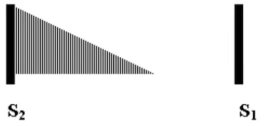
A 47-year-old man comes to the clinic for a routine check-up. Which of the following is most likely to be seen in this patient?
Show answer and explanation
Correct answer: B. Blood pressure of 150/50
The answer
There is no real vignette here, on purpose: the question wants you to name the murmur from the drawing. The murmur starts right after S2. Diastole runs from S2 to S1, so this is a diastolic murmur. It is loudest at the start and then fades, an early diastolic, decrescendo murmur. That is aortic regurgitation, not mitral stenosis, whose rumble comes later in diastole.
Aortic regurgitation causes a wide pulse pressure, meaning a big difference between the systolic and diastolic pressures. During diastole the aortic valve is closed, but it is incompetent. Blood falls back out of the aorta into the left ventricle, so the diastolic pressure collapses. That gives a reading like 150/50, which is option B.
Why A, C, D and E are wrong
A, angina, and E, pulsus parvus et tardus, are classic for aortic stenosis, a systolic murmur, not a diastolic one. C, cannon A waves, come from AV dissociation, such as third-degree heart block. D, myxomatous degeneration, is the exam buzzword for mitral valve prolapse.
High-yield tips
★ High-yield tip #1
The diastolic murmurs
When a murmur sits in diastole, ask yourself which diastolic murmurs you know. The two left-sided ones are aortic regurgitation and mitral stenosis. The right-sided ones, pulmonic regurgitation and tricuspid stenosis, are close to nonexistent on the USMLE; tricuspid stenosis is under 1% of valve disease. There are other unusual sounds, like a tumor plop from an atrial myxoma, or a venous hum.
★ High-yield tip #2
Describing the aortic regurgitation murmur
Aortic regurgitation is described as an early diastolic, decrescendo murmur, and sometimes as a decrescendo holodiastolic murmur. That last term can be confusing, because a holodiastolic murmur should fill all of diastole. Still, both descriptions mean the same murmur: aortic regurgitation.
★ High-yield tip #3
Mitral stenosis versus aortic regurgitation
Mitral stenosis sounds different. There is an opening snap shortly after S2, followed by a mid-to-late diastolic rumble. So the sound sits in the later part of the space between S2 and S1, not right after S2. Aortic regurgitation, by contrast, starts right at S2 and then fades.
★ High-yield tip #4
Spotting a wide pulse pressure
Look for any blood pressure with a big gap between the two numbers, like 120/40 or 150/50. In aortic regurgitation the diastolic number can fall extremely low. Whenever you see that gap, think of a leaky aortic valve.
★ High-yield tip #5
Causes of aortic regurgitation
A bicuspid aortic valve is a common cause of both aortic stenosis and aortic regurgitation. For aortic regurgitation, the cause the exam most wants you to know is aortic dissection. Hypertension or cocaine use can cause a dissection that spreads backward toward the aortic root, causing aortic root dilation so the valve can no longer close properly. Dissection causes aortic regurgitation, not aortic stenosis.
★ High-yield tip #6
Aortic stenosis
Angina and pulsus parvus et tardus are both classic for aortic stenosis. Aortic stenosis is a mid-systolic, crescendo-decrescendo murmur, so it sits between S1 and S2, not between S2 and S1. Remember SAD: syncope, angina and dyspnea. Pulsus parvus et tardus means a weak, slow-rising pulse, and the murmur radiates to the carotids.
★ High-yield tip #7
Cannon A waves
When students don’t know an answer, they often pick the strange-sounding option. You hardly need the jugular venous pulse curve for the USMLE. Cannon A waves happen when the right atrium contracts against a closed tricuspid valve. This occurs with AV dissociation, such as third-degree heart block, when the atria and ventricles are no longer in sync. The pressure bounces back up into the jugular vein.
★ High-yield tip #8
Myxomatous degeneration
On the USMLE, myxomatous degeneration is the buzzword for mitral valve prolapse, which gives a mid-systolic click. It is the term used for how connective tissue disorders like Marfan syndrome and Ehlers-Danlos affect the mitral valve. These disorders can also raise the risk of dissection and aortic regurgitation, but the exam ties myxomatous degeneration to mitral valve prolapse, not aortic regurgitation.
Sources: StatPearls: Aortic Regurgitation; StatPearls: Aortic Valve Disease; Clinical Methods: Diastolic Murmurs; StatPearls: Physiology, Heart Sounds; StatPearls: Mitral Stenosis; StatPearls: Aortic Stenosis; Clinical Methods: Systolic Murmurs; Clinical Methods: The Jugular Venous Pressure and Pulse Contour; StatPearls: Third-Degree Atrioventricular Block; StatPearls: Mitral Valve Prolapse; StatPearls: Aortic Dissection; StatPearls: Tricuspid Stenosis; StatPearls: Atrial Myxoma; StatPearls: Bicuspid Aortic Valve
Question 4
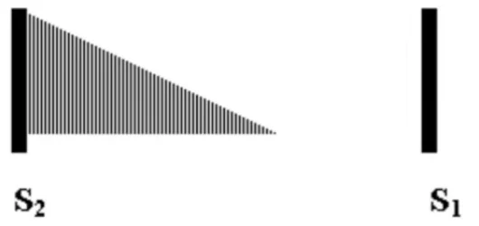
A 42-year-old man is examined in the emergency department. Cardiopulmonary auscultation is performed. Which of the following is most likely to be seen in this patient?
Show answer and explanation
Correct answer: A. Cocaine use
The answer
There is no vignette, on purpose: the clue is the murmur in the diagram. S1 marks the start of systole and S2 marks the start of diastole, so the gap between S2 and S1 is diastole. This murmur begins right at S2 and is clearly decrescendo. It does not fill all of diastole, but that does not matter: an early diastolic, decrescendo murmur is aortic regurgitation.
Cocaine causes hypertension, and hypertension can cause aortic dissection. A dissection can spread backward toward the aortic root, disrupting the aortic annulus and causing aortic regurgitation. Across these exams, aortic dissection is the most common cause of aortic regurgitation you will see.
Why B, C, D and E are wrong
B, repeated fleeting left-sided chest pain, describes mitral valve prolapse syndrome, which gives a mid-systolic click, not this murmur. C, a murmur that increases with inspiration, is right-sided, so tricuspid or pulmonic, but aortic regurgitation is left-sided. D, myocardial ischemia, can happen with cocaine, but it is not the best answer: ischemia classically relates to mitral regurgitation. E, myxomatous degeneration, is the histology of mitral valve prolapse, not aortic regurgitation.
High-yield tips
★ High-yield tip #1
The aortic regurgitation murmur
Aortic regurgitation is an early diastolic, decrescendo murmur, and it can also be described as holodiastolic, meaning present throughout diastole. Pulmonic regurgitation can sound similar, but it is virtually nonexistent on the USMLE, so an early diastolic, decrescendo murmur on the exam is far more likely to be aortic regurgitation.
★ High-yield tip #2
The hidden murmur in aortic dissection
The exam won’t make this easy. You might get a long, twelve-line vignette about a dissection that simply mentions a diastolic murmur and nothing else. Students recognize the dissection but miss the murmur. That diastolic murmur is aortic regurgitation.
★ High-yield tip #3
Mitral valve prolapse syndrome
Most people with mitral valve prolapse have no symptoms. A minority do, called mitral valve prolapse syndrome, with atypical chest pain that the exam describes as fleeting chest pain on the left. A Step 2 CK question might describe a 30-year-old woman with about 30 such episodes, whose father died of a heart attack. The answer is reassurance, with no specific treatment needed. Mitral valve prolapse gives a mid-systolic click between S1 and S2.
★ High-yield tip #4
Respiration and tricuspid regurgitation
Left-sided murmurs increase with expiration, and right-sided murmurs increase with inspiration. The exam uses this: a holosystolic murmur that increases with inspiration is tricuspid regurgitation. Causes include IV drug use, which leads to tricuspid vegetations, and carcinoid syndrome. Cor pulmonale, or pulmonary hypertension, also causes it: the right ventricle expands and dilates the tricuspid annulus.
★ High-yield tip #5
Ischemia and mitral regurgitation
Papillary muscle rupture after a heart attack is the highest-yield cause of mitral regurgitation on the USMLE, though it is not the most common cause in real life. Ischemic remodeling after a heart attack also causes it, and dilated cardiomyopathy stretches the mitral annulus, which also causes mitral regurgitation.
★ High-yield tip #6
Myxomatous degeneration
In Marfan syndrome and Ehlers-Danlos, histology can show a thickened, proteoglycan-rich valve extracellular matrix, called myxomatous degeneration. The exam ties it to mitral valve prolapse, with its mid-systolic click. Marfan syndrome can cause aortic regurgitation through aortic root dilation, or through cystic medial necrosis leading to aortic dissection. Myxomatous degeneration is the histology for mitral valve prolapse, not aortic regurgitation.
Sources: StatPearls: Aortic Dissection; StatPearls: Cocaine Toxicity; StatPearls: Aortic Regurgitation (Archived); MedlinePlus: Aortic Regurgitation; StatPearls: Physiology, Heart Sounds; StatPearls: Aortic Insufficiency; MedlinePlus: Mitral Valve Prolapse; StatPearls: Mitral Valve Prolapse; StatPearls: Tricuspid Regurgitation; StatPearls: Mitral Regurgitation; Review: TGF-beta Signaling in Myxomatous Mitral Valve Degeneration (Front Cardiovasc Med 2022); StatPearls: Marfan Syndrome; StatPearls: Ehlers-Danlos Syndromes; StatPearls: Tricuspid Valve Endocarditis; StatPearls: Carcinoid Syndrome; StatPearls: Papillary Muscle Rupture
Question 5
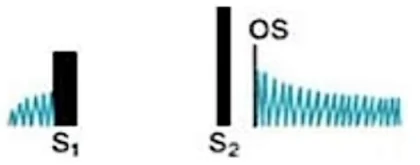
A 47-year-old woman is seen in the clinic. Cardiac auscultation is performed. Which of the following is the most likely explanation for this patient’s findings?
Show answer and explanation
Correct answer: C. Immune hypersensitivity
The answer
The clue is the diagram. S1 marks the start of systole and S2 the start of diastole. Between S1 and S2 there is no sound, so the murmur is not in systole. In diastole there is an opening snap, marked OS, followed by a mid-to-late diastolic rumble. That is mitral stenosis.
Worldwide, mitral stenosis is most often due to rheumatic heart disease, and that is an immune hypersensitivity reaction. After Streptococcus pyogenes pharyngitis, antibodies form against the M protein of group A strep. Those antibodies cross-react with antigens on the mitral valve and damage it. That is why the answer is C.
Why A, B, D and E are wrong
A, aortic dissection, can cause aortic regurgitation, an early diastolic murmur, but the start of diastole is silent here. B, a calcified bicuspid aortic valve, causes aortic stenosis, a systolic murmur. D, intravenous drug use, classically causes tricuspid regurgitation, also a systolic murmur. E, myxomatous degeneration, goes with mitral valve prolapse and a mid-systolic click. This diagram has no systolic sound at all.
High-yield tips
★ High-yield tip #1
Tricky wording for mitral stenosis
Be ready for tricky wording. A question may not mention an opening snap at all and just describe a rumbling diastolic murmur. That is also mitral stenosis. So when you read about a rumbling murmur in diastole, think of mitral stenosis even without the snap.
★ High-yield tip #2
Rheumatic heart disease, regurgitation then stenosis
Rheumatic heart disease changes the mitral valve over time. A child who gets rheumatic heart disease first develops mitral regurgitation. Over years, the valve scars and becomes stenotic. So a 12-year-old with acute rheumatic heart disease has mitral regurgitation, and years later, mitral stenosis.
★ High-yield tip #3
Other causes of mitral stenosis
Rheumatic heart disease is not the only cause of mitral stenosis. In developed countries, calcification of the mitral annulus in older adults is also a common cause. Rarer causes include lupus, which affects the valves through Libman-Sacks endocarditis.
★ High-yield tip #4
The aortic regurgitation murmur
Aortic regurgitation is an early diastolic murmur, also called a decrescendo holodiastolic or pan-diastolic murmur. It starts right at S2, the very beginning of diastole. So a murmur diagram with a silent start of diastole argues against aortic regurgitation.
★ High-yield tip #5
Aortic dissection and aortic regurgitation
For the USMLE, know that an aortic dissection can spread backward toward the aortic root, disrupting the aortic annulus and causing aortic regurgitation. It is the highest-yield cause of aortic regurgitation on the exam. Dissection can come from hypertension, cocaine use, or connective tissue disorders like Marfan syndrome and Ehlers-Danlos. In Marfan, the lesion behind it is cystic medial necrosis.
★ High-yield tip #6
Aortic stenosis and tricuspid regurgitation
A calcified bicuspid aortic valve points to aortic stenosis. That is a mid-systolic, crescendo-decrescendo murmur, with syncope, angina and dyspnea. Intravenous drug use classically causes tricuspid regurgitation, which is a holosystolic murmur that increases with inspiration. Both are systolic murmurs, heard between S1 and S2.
★ High-yield tip #7
Myxomatous degeneration
Myxomatous degeneration goes with mitral valve prolapse and its mid-systolic click. It describes the histology of proteoglycan deposits in the valve’s extracellular matrix, and it is seen with Marfan syndrome and Ehlers-Danlos. In one of these patients with a mid-systolic click, the answer is myxomatous degeneration.
★ High-yield tip #8
Telling them apart
If a patient with Marfan syndrome or Ehlers-Danlos instead has an aortic dissection, with sudden, severe, tearing chest or back pain, the answer is cystic medial necrosis. Mitral valve prolapse is the most common valve abnormality, so know it well: a mid-systolic click. Mitral stenosis is an opening snap in diastole, followed by a mid-to-late diastolic rumble.
Sources: StatPearls: Rheumatic Heart Disease; StatPearls: Mitral Stenosis; StatPearls: Acute Rheumatic Fever; StatPearls: Physiology, Heart Sounds; StatPearls: Libman-Sacks Endocarditis; StatPearls: Aortic Regurgitation (Archived); StatPearls: Aortic Stenosis; StatPearls: Tricuspid Valve Endocarditis; StatPearls: Tricuspid Regurgitation; StatPearls: Mitral Valve Prolapse; Review: TGF-beta Signaling in Myxomatous Mitral Valve Degeneration (Front Cardiovasc Med 2022); StatPearls: Marfan Syndrome; StatPearls: Ehlers-Danlos Syndromes; StatPearls: Aortic Dissection; ESC: Mitral Valve Incompetence, Epidemiology and Causes
Question 6
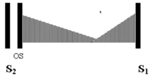
A 41-year-old woman is evaluated in the outpatient cardiology clinic. Her cardiac auscultation is shown. Which of the following is the most likely explanation for this patient’s findings?
Show answer and explanation
Correct answer: D. History of rheumatic heart disease
The answer
Read the diagram. S2 marks the start of diastole and S1 marks the start of systole. The murmur sits between S2 and S1, so it is diastolic. The giveaway is the opening snap, labeled OS, just after S2. Ask yourself which diastolic murmur comes with an opening snap. The answer is mitral stenosis.
The stiff valve snaps open a little way into diastole. Drawings of mitral stenosis vary in shape, but every one of them shows the opening snap followed by a diastolic murmur. And mitral stenosis is most often caused by rheumatic heart disease, which scars the mitral valve over the years. So the answer is D, a history of rheumatic heart disease.
Why A, B, C and E are wrong
A, a bicuspid aortic valve, causes aortic stenosis, a mid-systolic or crescendo-decrescendo murmur, but the stretch from S1 to S2 is blank. B, carcinoid syndrome, can cause tricuspid regurgitation, a holosystolic murmur, not a diastolic one. C, a dissection, can cause aortic regurgitation, an early diastolic murmur that begins right at S2, but here nothing starts at S2 and the shape dips down and rises again. E, myxomatous degeneration, is mitral valve prolapse, which gives a mid-systolic click.
High-yield tips
★ High-yield tip #1
How rheumatic heart disease causes mitral stenosis
Rheumatic heart disease follows strep throat in childhood, caused by Streptococcus pyogenes, which is group A strep. The immune system makes antibodies against the bacterium’s M protein, and these cross-react with mitral valve antigens. This is molecular mimicry. In the child, the acute result is mitral regurgitation, a holosystolic murmur. Over the years the valve scars, and later in life it becomes mitral stenosis.
★ High-yield tip #2
Causes of mitral stenosis
Rheumatic heart disease is the predominant cause of mitral stenosis worldwide, and on the exam you should link the two almost every time. Much rarer causes of mitral valve disease include Libman-Sacks endocarditis in lupus, with vegetations on both sides of the valve, and nonbacterial thrombotic endocarditis, also called marantic endocarditis, which can occur with cancer.
★ High-yield tip #3
Bicuspid valve in children
A bicuspid aortic valve does not have to show up as a valve that calcifies early in middle age. The exam can give you a child with aortic stenosis from a bicuspid valve, which throws some students off. For example, a three-month-old with a grade 4/6 mid-systolic murmur. The answer is still bicuspid aortic valve. It does not have to be an adult.
★ High-yield tip #4
Aortic stenosis
For aortic stenosis, remember SAD: syncope, angina and dyspnea. The stiffened left ventricle can sometimes produce an S4 heart sound. The pulse is pulsus parvus et tardus, a weak, slow-rising pulse, so the carotid pulse has a delayed upstroke.
★ High-yield tip #5
Carcinoid syndrome
Carcinoid syndrome means a carcinoid tumor, classically of the appendix or small bowel, that secretes serotonin. In some patients this leads to fibrous, plaque-like thickening of the tricuspid valve and tricuspid regurgitation. Picture a patient with flushing, a fast heart rate and diarrhea, with raised urine 5-HIAA, the main test used to diagnose carcinoid syndrome.
★ High-yield tip #6
Tricuspid regurgitation
Tricuspid regurgitation gives a holosystolic murmur that gets louder with inspiration. Besides carcinoid syndrome, it can come from endocarditis in people who inject drugs. But the highest-yield cause is pulmonary hypertension, or cor pulmonale. Pressure backs up into the right ventricle, which dilates and stretches the tricuspid annulus. So a patient with COPD and a holosystolic murmur that increases with inspiration has tricuspid regurgitation from pulmonary hypertension.
★ High-yield tip #7
Dissection and aortic regurgitation
An aortic dissection can extend backward toward the aortic root, disrupt the aortic annulus, and cause aortic regurgitation. That gives an early diastolic, decrescendo murmur that begins right at S2. Dissection is a high-yield cause of aortic regurgitation, for example with Marfan syndrome, Ehlers-Danlos syndrome, cystic medial necrosis of the aorta, or cocaine use.
★ High-yield tip #8
Signs of aortic regurgitation
In aortic regurgitation, look for head bobbing, bounding pulses and a wide pulse pressure, a big gap between systolic and diastolic pressure, such as 160/50. The diastolic pressure collapses because blood falls back from the aorta into the left ventricle.
★ High-yield tip #9
The click versus the snap
Myxomatous degeneration refers to mitral valve prolapse, which gives a mid-systolic click. When you first learn this, it is easy to mix up the mid-systolic click and the opening snap. The click is mitral valve prolapse. The opening snap is mitral stenosis. Also, mitral valve prolapse is not the same thing as mitral regurgitation.
★ High-yield tip #10
Marfan and the mitral valve
We said Marfan syndrome and Ehlers-Danlos syndrome can cause dissection with aortic regurgitation. They can also cause proteoglycan buildup in the mitral valve’s extracellular matrix, which leads to mitral valve prolapse. Myxomatous degeneration is a buzzword for mitral valve prolapse, not for aortic regurgitation. The exam may simply describe a man who is six foot nine, with long hands and flat feet, and a mid-systolic click.
★ High-yield tip #11
Mitral valve prolapse on the exam
Mitral valve prolapse is very common, found in about 2 to 3% of people, and most patients have no symptoms. When you see Marfan features and a mid-systolic click, the answer is simply myxomatous degeneration.
Sources: StatPearls: Mitral Stenosis; StatPearls: Rheumatic Heart Disease; StatPearls: Physiology, Heart Sounds; StatPearls: Aortic Stenosis; StatPearls: Acute Rheumatic Fever; StatPearls: Libman-Sacks Endocarditis; StatPearls: Carcinoid Syndrome; StatPearls: Tricuspid Regurgitation; StatPearls: Tricuspid Valve Endocarditis; StatPearls: Pulmonary Hypertension; StatPearls: Aortic Dissection; StatPearls: Aortic Regurgitation (Archived); StatPearls: Mitral Valve Prolapse; StatPearls: Surgical Approaches to Congenital Aortic Stenosis; StatPearls: Marfan Syndrome; StatPearls: Ehlers-Danlos Syndromes; StatPearls: Cocaine Toxicity; StatPearls: Aortic Insufficiency; StatPearls: Physiology, Pulse Pressure; Review: TGF-beta Signaling in Myxomatous Mitral Valve Degeneration (Front Cardiovasc Med 2022); MedlinePlus: Marfan Syndrome
Question 7
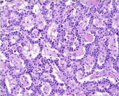
A 52-year-old man comes to the clinic for a 4-month history of intermittent palpitations, facial flushing, and diarrhea. Urinary 5-hydroxyindoleacetic acid is increased. A histology image is shown. Which of the following is most likely to be seen in this patient?
Show answer and explanation
Correct answer: B. 2/6 Holosystolic murmur that increases with inspiration
The answer
The stem hands you the diagnosis: a carcinoid tumor. It can cause flushing, palpitations and diarrhea, and raised urine 5-HIAA is characteristic. You do not need to know the image. It just shows basophilic cells. Basophilic means purple, and eosinophilic means pink.
Once a carcinoid tumor spreads to the liver, its serotonin reaches the right heart. There it lays down fibrous, plaque-like deposits on the right-sided valves, especially the tricuspid, so the valve leaks. Tricuspid regurgitation is a holosystolic murmur that increases with inspiration, which is option B.
Why A, C, D and E are wrong
A, an early diastolic murmur, is aortic regurgitation, or technically pulmonic regurgitation. C, a mid-systolic murmur, is aortic stenosis; pulmonic stenosis is less likely with carcinoid and would increase with inspiration. D, an opening snap with a diastolic murmur, is mitral stenosis. E, a to-and-fro murmur, is a systolic plus a separate diastolic murmur, not the classic carcinoid murmur.
High-yield tips
★ High-yield tip #1
Carcinoid tumors and the liver
Carcinoid tumors occur in the GI tract, such as the small bowel and appendix, and in the lungs as bronchial carcinoids. They secrete serotonin, and its urine breakdown product, 5-HIAA, is high yield. The liver normally breaks these hormones down. If the tumor spreads to the liver, the hormones reach the circulation directly, and that is when the right-sided valves get damaged, most often the tricuspid.
★ High-yield tip #2
Inspiration and right-sided murmurs
A holosystolic murmur that increases with inspiration is tricuspid regurgitation, and you have to know that. Right-sided murmurs get louder with inspiration because inspiration increases venous return to the right heart. If a holosystolic murmur is loudest at the apex and radiates to the left axilla, and the question does not mention inspiration, think mitral regurgitation instead.
★ High-yield tip #3
Causes of tricuspid regurgitation
It sounds odd, but the exam wants you to know that carcinoid tumors can cause tricuspid regurgitation. Other causes include right-sided endocarditis in people who inject drugs, and cor pulmonale. In pulmonary hypertension, pressure backs up into the right heart and stretches the tricuspid annulus, so the valve can no longer close.
★ High-yield tip #4
Aortic regurgitation
An early diastolic murmur points to aortic regurgitation. Technically it could be pulmonic regurgitation, since both are described as early diastolic, decrescendo murmurs. For aortic regurgitation, think head bobbing, bounding pulses and a wide pulse pressure, a big gap between systolic and diastolic pressure. A high-yield cause is a type A aortic dissection that extends back to the aortic root and damages the valve.
★ High-yield tip #5
Mid-systolic murmurs and carcinoid
A mid-systolic murmur means aortic stenosis, or possibly pulmonic stenosis. Pulmonic stenosis can occur with carcinoid tumors, but it is less likely than tricuspid regurgitation. If the exam wants pulmonic stenosis, it describes a mid-systolic murmur that increases with inspiration.
★ High-yield tip #6
Aortic stenosis
Aortic stenosis gives a crescendo-decrescendo murmur. Look for pulsus parvus et tardus, a slow-rising pulse with a delayed carotid upstroke, and SAD: syncope, angina and dyspnea. A bicuspid aortic valve is a high-yield cause. Congenital aortic stenosis does not need heavy calcification, and it can present in children too, which confuses students who assume it has to be an adult.
★ High-yield tip #7
Mitral stenosis
An opening snap with a diastolic murmur is mitral stenosis, and on the exam, mitral stenosis means a history of rheumatic heart disease. In young people, rheumatic heart disease most often shows up as mitral regurgitation. Over the following decades the valve scars and can become stenotic. So the classic finding is an opening snap followed by a low-pitched diastolic rumble.
★ High-yield tip #8
Mitral stenosis in pregnancy
The exam can also give a pregnant woman with a diastolic murmur and shortness of breath from the second trimester onward. That is mitral stenosis becoming symptomatic. Plasma volume rises by about 45 to 55% in pregnancy, and hemodynamic demands peak in the second trimester. Stenosis that was silent before now causes symptoms. The cause is still a history of rheumatic heart disease. Pregnancy simply unmasks it.
★ High-yield tip #9
To-and-fro versus continuous murmurs
A to-and-fro murmur is a systolic murmur plus a separate diastolic murmur, as when aortic stenosis and aortic regurgitation occur together. Don’t confuse it with a continuous, machinery-like murmur, which begins in systole and continues past S2. Neither one is the classic murmur of carcinoid heart disease, which most often causes tricuspid regurgitation.
★ High-yield tip #10
PDA wording on the exam
Exam questions sometimes use to-and-fro loosely for a PDA, whose classic murmur is continuous and machinery-like. One offline NBME form for Step 2 CK reportedly gives congenital rubella syndrome with a to-and-fro murmur, meaning a PDA, so know that rubella causes it. Continuous murmurs can also come from outside the heart, such as an arteriovenous fistula at the arterial puncture site after cardiac catheterization, which gives a continuous bruit.
Sources: StatPearls: Tricuspid Regurgitation; StatPearls: Carcinoid Syndrome; StatPearls: Physiology, Heart Sounds; StatPearls: Hepatojugular Reflux; MedlinePlus: Carcinoid syndrome; MedlinePlus: 5-HIAA urine test; StatPearls: Histology, Staining; StatPearls: Gastrointestinal Neuroendocrine Tumors; PDQ: Gastrointestinal Neuroendocrine Tumors Treatment (Patient); StatPearls: Mitral Regurgitation; StatPearls: Physiology, Cardiovascular Murmurs; StatPearls: Aortic Valve Disease; StatPearls: Aortic Stenosis; StatPearls: Mitral Stenosis; StatPearls: Rheumatic Heart Disease; StatPearls: Tetralogy of Fallot; StatPearls: Left Ventricular Outflow Tract Obstruction; StatPearls: Balloon Valvuloplasty; StatPearls: Physiology, Pregnancy; StatPearls: Cardiac Disease in Pregnancy; StatPearls: Patent Ductus Arteriosus; Rubella, Congenital Syndrome (CRS) 2010 Case Definition; StatPearls: Cardiac Catheterization Risks and Complications
Question 8
A 16-year-old boy has a murmur discovered during a preseason physical examination for his school soccer team. Physical examination shows a systolic murmur loudest over the mid-sternal area at the 2nd intercostal space. There is no change in the murmur with Valsalva. He is 6’5″ and 200 pounds. Vital signs are normal. Which of the following is the most likely cause of this patient’s cardiac findings?
Show answer and explanation
Correct answer: C. Bicuspid aortic valve
The answer
The key clues are a systolic murmur loudest over the mid-sternal area at the 2nd intercostal space, in a 16-year-old, with no change on Valsalva. A systolic murmur there fits a stenotic aortic valve, and in someone this young the likely cause is a bicuspid aortic valve, option C. No change with Valsalva fits too: Valsalva lowers preload, so most murmurs, aortic stenosis included, get softer or stay the same.
Why A and B are wrong
A, asymmetric septal hypertrophy, is hypertrophic obstructive cardiomyopathy, whose murmur gets louder with Valsalva. B, an atrial septal defect, would be described with fixed splitting of S2.
Why D and E are wrong
D, cystic medial necrosis, leads to aortic dissection and aortic regurgitation, a diastolic murmur, but this murmur is systolic. His height of 6’5″ hints at Marfan syndrome on purpose, but people with Marfan syndrome are usually tall and slender, and he weighs 200 pounds. E, myxomatous degeneration, is mitral valve prolapse, with a mid-systolic click and a murmur that gets worse with Valsalva.
High-yield tips
★ High-yield tip #1
Who gets a bicuspid aortic valve
Most people with a bicuspid aortic valve do not have Turner syndrome. It can occur on its own or run in families, where the pattern can be autosomal dominant.
★ High-yield tip #2
Bicuspid valve in children
Symptoms of stenosis from a bicuspid valve typically begin between ages 50 and 70, but many students wrongly learn that a bicuspid valve only causes trouble in adulthood. The exam will happily give you a bicuspid valve in children, even in an infant a few months old, and almost everyone is surprised.
★ High-yield tip #3
Valsalva and murmurs
Valsalva sharply raises intrathoracic pressure and reduces preload, so less blood fills the heart. Whenever a vignette mentions Valsalva, think less volume in the heart. With less volume, most murmurs, aortic stenosis included, get softer. The point is that they do not get louder, so a murmur that does not change fits too. The exceptions are HOCM and mitral valve prolapse, which get worse. Going from lying down to standing works the same way.
★ High-yield tip #4
Splitting of S2
B, an atrial septal defect, would be described with fixed splitting of S2. Some students ask why vignettes don’t always say wide fixed splitting. Wide splitting means the pulmonic valve closes late, as with right-sided overload. Paradoxical splitting means the aortic valve closes late, as with left bundle branch block or severe aortic stenosis. So the key word for an atrial septal defect is fixed, and vignettes may simply say fixed splitting.
★ High-yield tip #5
Cystic medial necrosis
Cystic medial necrosis describes degenerative changes in the wall of the aorta, classically in Marfan syndrome, though it also occurs with hypertension. It sets the stage for aortic dissection, and a dissection reaching the aortic root causes aortic regurgitation, a diastolic murmur.
★ High-yield tip #6
Aortic regurgitation versus stenosis
The murmur of aortic regurgitation is a decrescendo diastolic murmur, which can be holodiastolic. It is loudest right after S2, so it is also called early diastolic. One important cause of aortic regurgitation is aortic dissection reaching the aortic annulus. Look for bounding pulses, head bobbing, and a wide pulse pressure. Aortic stenosis instead gives pulsus parvus et tardus, slow-rising pulses, and its murmur can radiate to the carotids.
★ High-yield tip #7
Myxomatous degeneration
E, myxomatous degeneration, is the valve change behind mitral valve prolapse. It can occur on its own or with connective tissue disorders such as Ehlers-Danlos syndrome and Marfan syndrome. You would expect a mid-systolic click, and the murmur gets louder with Valsalva.
★ High-yield tip #8
Vague murmur locations
Exam questions can be vague about murmur location and may not say right or left. Sometimes they place an aortic valve murmur at the left sternal border, second intercostal space, which is technically the pulmonic area, not the aortic area.
Sources: StatPearls: Bicuspid Aortic Valve; StatPearls: Surgical Approaches to Congenital Aortic Stenosis; StatPearls: Physiology, Cardiovascular Murmurs; StatPearls: Aortic Stenosis; StatPearls: Valsalva Maneuver; Clinical Methods: The Second Heart Sound; StatPearls: Aortic Dissection; StatPearls: Aortic Regurgitation; MedlinePlus Genetics: Marfan syndrome; StatPearls: Mitral Valve Prolapse
Question 9
A 17-year-old boy is brought to the physician after the school nurse at his high school heard a murmur. He has no past medical history. Blood pressure is 130/40. Which of the following is most likely to be seen in this patient?
Show answer and explanation
Correct answer: B. 2/6 decrescendo holo-diastolic murmur
The answer
The key clue is the blood pressure of 130/40. That big gap between the systolic and diastolic pressures is called a wide pulse pressure, and it points to aortic regurgitation. In aortic regurgitation, systolic pressure rises and diastolic pressure falls, which widens that gap.
In aortic regurgitation, the leaky aortic valve lets blood flow back into the left ventricle during diastole. That backflow is heard as a decrescendo holodiastolic murmur, which is option B, and it is the murmur that goes with this boy’s wide pulse pressure.
Why A, C, D and E are wrong
A, a decrescendo mid-to-late diastolic murmur, points to mitral stenosis. C, a holosystolic murmur that increases with inspiration, is tricuspid regurgitation. D, a mid-systolic click, is mitral valve prolapse. E, a mid-systolic murmur with an ejection click, is aortic stenosis. None of them explains this boy’s wide pulse pressure.
High-yield tips
★ High-yield tip #1
How the aortic regurgitation murmur is described
Know the different ways a question can describe the aortic regurgitation murmur. It can be called a decrescendo holodiastolic murmur, an early diastolic murmur, or a murmur loudest immediately after S2, because diastole begins after S2. All of these wordings point to aortic regurgitation.
★ High-yield tip #2
Signs and causes of aortic regurgitation
The classic signs of aortic regurgitation are a wide pulse pressure, head bobbing, and bounding pulses. One important cause is aortic dissection spreading back toward the aortic root, for example with hypertension, Marfan syndrome or Ehlers-Danlos syndrome. A question does not have to give a cause; the wide pulse pressure alone can be enough.
★ High-yield tip #3
Pulmonic regurgitation
Pulmonic regurgitation would in theory give the same kind of murmur, but it would be described as getting louder with inspiration, like other right-sided murmurs. Its classic form, the Graham Steell murmur, comes from pulmonary hypertension stretching the pulmonic valve ring.
★ High-yield tip #4
Mitral stenosis and rheumatic heart disease
Mitral stenosis gives an opening snap shortly after S2, then a mid-to-late diastolic rumble. Worldwide, it is most often due to rheumatic heart disease. In young people, rheumatic heart disease most often shows up as mitral regurgitation, while symptoms of rheumatic mitral stenosis typically begin in the third or fourth decade.
★ High-yield tip #5
Mitral stenosis in pregnancy
Another classic vignette is a pregnant woman in her second or third trimester who becomes short of breath and has a diastolic murmur. That is mitral stenosis. Mild mitral stenosis may first cause symptoms in pregnancy, because plasma volume and cardiac output rise, and cardiac output peaks by mid-pregnancy.
★ High-yield tip #6
Causes of tricuspid regurgitation
Tricuspid regurgitation gives a holosystolic murmur that increases with inspiration. Its most common cause is secondary, from a right ventricle enlarged by pressure or volume overload, as in pulmonary hypertension and cor pulmonale, so lung disease can lead to it. A loud P2 is usually the first exam finding of pulmonary hypertension. Infective endocarditis in people who inject drugs is a primary cause.
★ High-yield tip #7
Mitral valve prolapse
Mitral valve prolapse gives a mid-systolic click and is one of the most common valve abnormalities. Most cases are primary, with no underlying connective tissue disease, although it can come with Ehlers-Danlos syndrome or Marfan syndrome. Most patients have no symptoms.
★ High-yield tip #8
Symptomatic mitral valve prolapse
Some patients do have symptoms, such as atypical chest pain or palpitations, without significant regurgitation. Picture a young woman with fleeting left-sided chest pain and a mid-systolic click. Once other causes are ruled out, management focuses on reassurance, lifestyle changes and treating symptoms, not on fixing the valve.
★ High-yield tip #9
Ejection click versus mid-systolic click
Do not confuse two different clicks. An ejection click is not the same thing as the mid-systolic click of mitral valve prolapse. An ejection click can be described in aortic stenosis vignettes, together with a mid-systolic murmur.
★ High-yield tip #10
Aortic stenosis
Aortic stenosis can cause syncope, angina and dyspnea, and pulsus parvus et tardus, meaning slow-rising pulses, also described as a slow carotid upstroke. Contrast aortic regurgitation, with bounding pulses, head bobbing and a wide pulse pressure. In patients under 70, the most common cause of aortic stenosis is a bicuspid aortic valve, which can run in families in an autosomal dominant pattern.
★ High-yield tip #11
Aortic stenosis in children
A bicuspid valve is not only about Turner syndrome, and it absolutely occurs in children. A pediatrics question might show a three-month-old with a mid-systolic murmur and an ejection click, and many students skip aortic stenosis because they think a bicuspid valve must be an adult problem. It does not have to calcify later in life first. Aortic stenosis can be the answer in a child.
Sources: StatPearls: Aortic Regurgitation; StatPearls: Physiology, Pulse Pressure; StatPearls: Mitral Stenosis; StatPearls: Rheumatic Heart Disease; StatPearls: Tricuspid Regurgitation; StatPearls: Pulmonary Hypertension; StatPearls: Mitral Valve Prolapse; StatPearls: Surgical Approaches to Congenital Aortic Stenosis; StatPearls: Aortic Dissection; StatPearls: Pulmonic Regurgitation; StatPearls: Physiology, Pregnancy; StatPearls: Aortic Stenosis; StatPearls: Bicuspid Aortic Valve
Question 10
A 26-year-old man visits a primary care clinic for a routine physical examination. He is 6′5″ and weighs 170 lbs. Cardiac auscultation shows a 2/6 midsystolic click. Physical examination shows long fingers, dental malocclusion, joint hypermobility, and scoliosis. Which of the following is the most likely cause of this patient’s cardiac finding?
Show answer and explanation
Correct answer: C. Myxomatous degeneration
The answer
The physical findings point to Marfan syndrome: a tall, thin build, long fingers, joint hypermobility, scoliosis and dental malocclusion. The 2/6 midsystolic click is the sign of mitral valve prolapse.
For the exam, myxomatous degeneration and mitral valve prolapse go together. The mitral leaflets degenerate: their structural collagen breaks down and becomes disorganized, and proteoglycans build up inside the leaflet, so the leaflets become thick and redundant. There is nothing to overanalyze here.
Why A, B, D and E are wrong
A, a bicuspid aortic valve, points to aortic stenosis, a crescendo-decrescendo ejection murmur. B, incompetence of the aortic valve leaflets, is aortic regurgitation, a decrescendo murmur in early diastole. D, narrowing of the mitral orifice, is mitral stenosis, which gives an opening snap in diastole. E, rupture of the chordae tendineae, causes mitral regurgitation, a holosystolic murmur. None of them gives a midsystolic click.
High-yield tips
★ High-yield tip #1
Connective tissue disorders
Mitral valve prolapse and aortic regurgitation are both seen in connective tissue disorders such as Marfan syndrome and Ehlers-Danlos syndrome. In a patient with Marfan features, the type of murmur tells you which lesion is present.
★ High-yield tip #2
The click versus the opening snap
Do not confuse a midsystolic click with an opening snap. An opening snap is heard in diastole and points to mitral stenosis. Think of it when a patient has a history of rheumatic fever. Most mitral stenosis is a late result of rheumatic heart disease from rheumatic fever in childhood.
★ High-yield tip #3
The Marfan trap, aortic regurgitation
Because aortic regurgitation is also seen in Marfan syndrome and Ehlers-Danlos, it is a tempting trap when a question describes Marfan features. That link is true, but aortic regurgitation would not cause a midsystolic click. It causes a high-pitched, blowing decrescendo murmur that starts in early diastole.
★ High-yield tip #4
Bicuspid aortic valve
A bicuspid aortic valve points to aortic stenosis, which gives a midsystolic, crescendo-decrescendo ejection murmur, not a midsystolic click. A bicuspid aortic valve is classic for Turner syndrome, but it can also simply run in families, sometimes in an autosomal dominant pattern.
★ High-yield tip #5
Mitral regurgitation after a heart attack
Rupture of the chordae tendineae causes mitral regurgitation. The classic exam link is a myocardial infarction: ischemia damages the papillary muscles that anchor the chordae, and papillary muscle rupture causes acute mitral regurgitation. That gives a holosystolic murmur that classically radiates to the axilla, not a midsystolic click.
Sources: StatPearls: Mitral Valve Prolapse; MedlinePlus Genetics: Marfan syndrome; StatPearls: Marfan Syndrome; StatPearls: Aortic Insufficiency; StatPearls: Physiology, Heart Sounds; StatPearls: Aortic Stenosis; StatPearls: Turner Syndrome; StatPearls: Bicuspid Aortic Valve (Archived); StatPearls: Mitral Stenosis; StatPearls: Mitral Regurgitation; StatPearls: Rheumatic Heart Disease; StatPearls: Acute Rheumatic Fever
Question 11
A 15-year-old boy is brought to the clinic by his mother for a six-month history of shortness of breath when running. He appears healthy and does not have any symptoms of chest pain, syncope, or palpitations. His vitals are: temperature 98.6°F, HR 74, RR 16, BP 118/76. Cardiac examination shows a blowing diastolic murmur at the right upper sternal border. Radial and pedal pulses are 3+. What’s the most likely mechanism for these findings?
Show answer and explanation
Correct answer: E. Left ventricular volume overload
The answer
This question comes down to one thing: do you recognize a blowing diastolic murmur at the right upper sternal border? Only a handful of murmurs are diastolic, and here they are describing aortic regurgitation.
The right upper sternal border is the aortic area, so a diastolic murmur there means aortic regurgitation. Blood that leaks back into the left ventricle during diastole adds to its filling, so aortic regurgitation is a preload problem, a volume overload. The 3+ radial and pedal pulses, meant here as brisk, strong pulses, fit too. That matches E, left ventricular volume overload.
Why A, B, C and D are wrong
A, increased cardiac output, is not the mechanism: the diastolic murmur points to a leaking aortic valve. B, increased pulmonary vascular resistance, fits pulmonary hypertension or cor pulmonale, right heart failure from lung disease. C, increased systemic vascular resistance, fits conditions such as systemic hypertension, but this blood pressure is a healthy 118/76. D, left ventricular pressure overload, points to aortic stenosis, a systolic murmur.
High-yield tips
★ High-yield tip #1
The diastolic murmurs
The word blowing is not the critical part. When you see diastolic, ask how many things it can be: aortic regurgitation, pulmonic regurgitation, or in theory mitral or tricuspid stenosis. The right upper sternal border is the aortic area, so a diastolic murmur there means aortic regurgitation, a decrescendo murmur that can last through diastole.
★ High-yield tip #2
Bounding pulses and pulse grading
Pulse grading is subjective. On the 0 to 4 scale used by StatPearls, 3+ is a normal, easily felt pulse and 4+ is bounding, so when a question gives 3+ as a clue, lean on the murmur, not that number. Aortic regurgitation classically gives a bounding, water-hammer pulse and can widen the pulse pressure, such as 120/40.
★ High-yield tip #3
Pressure overload versus volume overload
D, left ventricular pressure overload, is high yield. It points to aortic stenosis, and it can also mean systemic hypertension or the outflow obstruction of hypertrophic obstructive cardiomyopathy. Remember the pair: pressure overload, aortic stenosis; volume overload, aortic regurgitation. Aortic stenosis would give a midsystolic, crescendo-decrescendo systolic murmur, not a diastolic one.
★ High-yield tip #4
Don’t pigeonhole
Some students get thrown off by the word blowing because they expected a different description. The lesson is not to pigeonhole. Do not read one description in a textbook and decide a disease must always present that way. Many exam questions are not textbook, and if one is, consider yourself lucky.
Sources: StatPearls: Aortic Regurgitation (Archived); StatPearls: Aortic Insufficiency; StatPearls: Physiology, Cardiovascular Murmurs; StatPearls: Left Ventricular Hypertrophy; StatPearls: Physiology, Heart Sounds; StatPearls: Peripheral Pulse; StatPearls: Cor Pulmonale (Archived); StatPearls: Hypertensive Heart Disease; Vital signs; StatPearls: Hypertrophic Cardiomyopathy; StatPearls: Aortic Stenosis; StatPearls: Water Hammer Pulse; StatPearls: Physiology, Pulse Pressure
Question 12
A 47-year-old man visits his primary care physician for a routine physical examination. A 2/6 holosystolic murmur that increases in intensity with expiration is auscultated diffusely across the left hemithorax. What’s the most likely diagnosis?
Show answer and explanation
Correct answer: B. Mitral regurgitation
The answer
The key clue is a holosystolic murmur that increases in intensity with expiration. Expiration makes left-sided murmurs louder, so this is a left-sided holosystolic murmur, and that is mitral regurgitation, option B.
Here is why. When you breathe in, pressure inside the chest falls, which increases venous return to the right heart, so right-sided murmurs get louder. At the same time, the expanding lungs hold more blood in the pulmonary vessels, which briefly reduces flow from the pulmonary veins to the left heart. So inspiration softens left-sided murmurs, and expiration does the opposite: left-sided murmurs get louder.
Why A, C, D and E are wrong
A, aortic regurgitation, and C, pulmonic regurgitation, are diastolic murmurs, not systolic. D, tricuspid regurgitation, is holosystolic but right-sided, so it gets louder with inspiration. E, a ventricular septal defect, is also holosystolic, but getting louder with expiration is not how it is classically described.
High-yield tips
★ High-yield tip #1
Mitral versus tricuspid regurgitation
A holosystolic murmur that gets louder with expiration is mitral regurgitation. If the question instead said a holosystolic murmur that gets louder with inspiration, that would be tricuspid regurgitation.
★ High-yield tip #2
The three classic VSD presentations
A ventricular septal defect is also a holosystolic murmur. On exams it shows up in three classic ways, each a holosystolic murmur at the left sternal border: first, with a palpable thrill; second, with a diastolic rumble; third, with left atrial enlargement, a sign of a large shunt. That is how a VSD appears in vignettes.
★ High-yield tip #3
Aortic and pulmonic regurgitation
Pulmonic regurgitation and aortic regurgitation are both diastolic murmurs, heard in diastole, not systole. The aortic valve is on the left side of the heart, so aortic regurgitation gets louder with expiration. The pulmonic valve is on the right side, so pulmonic regurgitation gets louder with inspiration.
Sources: StatPearls: Mitral Regurgitation; StatPearls: Cardiogenic Pulmonary Edema; StatPearls: Pulsus Paradoxus; StatPearls: Hepatojugular Reflux; StatPearls: Physiology, Heart Sounds; StatPearls: Pulmonic Regurgitation; StatPearls: Ventricular Septal Defect; PMC review: Ventricular septal defect; StatPearls: Postinfarction Ventricular Septal Rupture
Question 13
A 50-year-old man is seen in clinic. He has a 2/6 decrescendo holodiastolic murmur auscultated down the right sternal border. Which of the following might also be seen in this patient?
Show answer and explanation
Correct answer: A. Blood pressure of 120/40
The answer
The clue is the murmur in the stem: a decrescendo holodiastolic murmur heard down the right sternal border. The first step is recognizing it as aortic regurgitation, which is classically a decrescendo diastolic murmur.
Choice A, a blood pressure of 120/40, is a wide pulse pressure, and it is the answer. The left ventricle pumps blood into the aorta, then, through the incompetent aortic valve, blood quickly falls back into the left ventricle. So the diastolic pressure falls steeply. That is why we get bounding pulses.
This question is not too complicated. You just have to identify the murmur in the vignette as aortic regurgitation, then remember that bounding pulses and a wide pulse pressure are classic for it.
Why B, C, D and E are wrong
B, flow across the interventricular septum, describes a ventricular septal defect, which gives a holosystolic murmur, not a diastolic one. C, a narrow aortic valve orifice, is aortic stenosis, a systolic murmur. D, systemic hypertension, is one of the causes of chronic aortic regurgitation, but it is not a finding this murmur predicts. E, tricuspid insufficiency, would be a holosystolic murmur.
High-yield tips
★ High-yield tip #1
How the aortic regurgitation murmur is described
A question may call the aortic regurgitation murmur a holodiastolic murmur loudest in early diastole, or simply a holodiastolic murmur. Classically it is decrescendo, loudest in early diastole and then fading. Any of these descriptions should make you think of aortic regurgitation.
★ High-yield tip #2
Murmur location is not always textbook
The classic aortic listening area is the second intercostal space at the right sternal border. A question does not have to say that. Some questions say the murmur radiates down the left or the right sternal border. Murmur questions are not always textbook.
★ High-yield tip #3
Radiation patterns
There are classic radiation patterns, such as mitral regurgitation radiating to the axilla or aortic stenosis radiating to the carotids, but a question need not mention them.
★ High-yield tip #4
Bounding pulses
Bounding pulses and head bobbing are characteristic of aortic regurgitation. A wide pulse pressure matters for aortic regurgitation, but it is not limited to it. Bounding pulses also occur with arteriovenous fistulas and sometimes with patent ductus arteriosus.
★ High-yield tip #5
Do not overlook the blood pressure
So remember: aortic regurgitation, wide pulse pressure, bounding pulses, a big gap between systolic and diastolic pressure. Some questions give a blood pressure like 160/60. Students often overlook it until someone points out the large difference between the two numbers.
★ High-yield tip #6
Three ways to describe a VSD
Exam questions describe a VSD with a holosystolic murmur at the left sternal border, or lower left sternal border, plus one of three extras: a palpable thrill, a diastolic rumble, or left atrial enlargement from a large shunt.
★ High-yield tip #7
Aortic stenosis
A narrow aortic valve orifice means aortic stenosis. It obstructs outflow from the left ventricle and gives a systolic crescendo-decrescendo murmur at the right upper sternal border, often radiating to the carotids.
★ High-yield tip #8
Hypertension and the S4
Systemic hypertension puts afterload on the left ventricle, and long-standing hypertension is better linked to an S4 heart sound. An S4 means the left ventricle is stiff. The thickened ventricle fills poorly in diastole, and the atrium contracting against it produces the S4. That diastolic dysfunction is classic for advanced hypertension.
★ High-yield tip #9
The tricuspid murmur
Tricuspid insufficiency would be a holosystolic murmur, classically at the left sternal border. The tricuspid listening area is the fourth intercostal space at the lower left sternal border.
Sources: StatPearls: Aortic Regurgitation (Archived); StatPearls: Aortic Insufficiency; StatPearls: Physiology, Pulse Pressure; StatPearls: Water Hammer Pulse; StatPearls: Physiology, Cardiovascular Murmurs; StatPearls: Ventricular Septal Defect; StatPearls: Aortic Stenosis; StatPearls: Hypertensive Heart Disease; StatPearls: Mitral Regurgitation; PMC review: Ventricular septal defect; StatPearls: Postinfarction Ventricular Septal Rupture; StatPearls: Physiology, Heart Sounds
Question 14
A 64-year-old woman comes to her primary care clinic because of hoarseness of voice for the past three months. She has a history of cardiovascular disease. A barium swallow shows indentation and posterior displacement of the esophagus. What is the most likely diagnosis?
Show answer and explanation
Correct answer: E. Ortner syndrome
The answer
The barium swallow finding looks obscure, but this question is really one anatomy fact. An enlarged left atrium can press on the esophagus, which explains the indentation and posterior displacement, and it can cause dysphagia. It can also press on the left recurrent laryngeal nerve, which causes hoarseness.
Hoarseness from left atrial enlargement pressing on the left recurrent laryngeal nerve is called Ortner syndrome, also known as cardiovocal syndrome. For the USMLE, just remember that an enlarged left atrium can cause both dysphagia and a hoarse voice.
Why A, B, C and D are wrong
A, Caplan syndrome, is rheumatoid arthritis plus pneumoconiosis. B, Felty syndrome, is rheumatoid arthritis plus neutropenia plus splenomegaly. C, Heyde syndrome, is aortic stenosis plus bleeding from angiodysplasia. D, Leriche syndrome, is aortoiliac atherosclerosis causing claudication and erectile dysfunction. None of them explains hoarseness and a displaced esophagus from an enlarged left atrium.
High-yield tips
★ High-yield tip #1
Caplan syndrome and the lungs in RA
Caplan syndrome is rheumatoid arthritis plus pneumoconiosis. Rheumatoid arthritis itself can cause restrictive lung disease, called rheumatoid lung. Methotrexate, the first-line DMARD for rheumatoid arthritis, can also injure the lungs and cause interstitial lung disease.
★ High-yield tip #2
Pneumoconiosis
Pneumoconiosis means lung disease from workplace dust, such as coal workers’ pneumoconiosis, silicosis and berylliosis. These are usually restrictive. So a person with rheumatoid arthritis who also has a dust exposure, such as working in a stone quarry with silica, fits Caplan syndrome. Caplan is spelled with a C.
★ High-yield tip #3
Felty syndrome
Felty syndrome is rheumatoid arthritis plus neutropenia plus splenomegaly. You don’t need to know why the three go together, only the combination. It looks low yield, but it does show up on Step 2 CK questions, where the answer is simply Felty syndrome.
★ High-yield tip #4
Heyde syndrome
Heyde syndrome is aortic stenosis plus bleeding from angiodysplasia. Angiodysplasia means dilated, tortuous small vessels in the wall of the bowel. It causes painless rectal bleeding in older adults, and it is one of the common causes in this age group, along with diverticular bleeding and colorectal cancer.
★ High-yield tip #5
Why aortic stenosis bleeds
Why the link? High shear stress across the narrowed aortic valve breaks up large von Willebrand factor multimers, so the fragile vessels bleed more easily. On Step 2 CK, a vignette may describe an older man who gets chest pain while arguing with his wife and also has a history of rectal bleeding. That is Heyde syndrome.
★ High-yield tip #6
Leriche syndrome
Leriche syndrome is atherosclerosis of the aortoiliac vessels in someone with advanced disease. It causes claudication in the buttocks and thighs, which reflects that high level of blockage, plus erectile dysfunction in men. If a man only gets calf pain when he walks, the disease is lower down, so that is not Leriche syndrome.
★ High-yield tip #7
Leriche syndrome on the exam
A classic question describes a man with advanced diabetes, a major risk factor for atherosclerosis, who has erectile dysfunction and no nocturnal erections. Absent nocturnal erections point to a physical cause rather than a psychological one, which fits Leriche syndrome.
Sources: StatPearls: Left Atrial Enlargement; PMC review: A Rare Case of a 54-year-old Male with Vocal Cord Paralysis Secondary to Left Atrial Enlargement; StatPearls: Thoracic Aorta Aneurysm; StatPearls: Caplan Syndrome; StatPearls: Restrictive Lung Disease; StatPearls: Rheumatoid Arthritis; StatPearls: Interstitial Lung Disease; StatPearls: Felty Syndrome; StatPearls: Heyde Syndrome; StatPearls: Angiodysplasia; StatPearls: Rectal Bleeding; StatPearls: Aortoiliac Occlusive Disease; StatPearls: Infrainguinal Occlusive Disease; StatPearls: Erectile Dysfunction
Question 15
A 68-year-old woman collapses and dies while playing tennis. She had no history of coronary artery disease or cardiac risk factors. Autopsy shows concentric left ventricular hypertrophy and a valvular defect. Which of the following valvular abnormalities is most likely related to her sudden death?
Show answer and explanation
Correct answer: B. Aortic stenosis
The answer
There isn’t much to go on: an older woman dies suddenly during physical activity, and the autopsy shows concentric left ventricular hypertrophy. Even if you don’t know where to start, know this: concentric hypertrophy reflects pressure overload on the left ventricle, meaning increased afterload. That points straight to aortic stenosis.
Sudden death can happen in aortic stenosis. The outflow tract of the left ventricle is narrowed, so during physical activity the left heart can be put under acute strain, which may set off a dangerous ventricular arrhythmia. The exact mechanism is not fully understood, but this is the likely explanation.
The heavy pressure load in aortic stenosis can also cause myocardial ischemia. If she had coronary artery disease, an acute ischemic event would be a possible mechanism. Because she had none, an arrhythmia from acute strain during exercise is the better explanation.
Why A, C, D, E, F, G and H are wrong
A, aortic regurgitation, and C, mitral regurgitation, are volume overloads that cause eccentric, not concentric, hypertrophy. D, mitral stenosis, raises pressure behind the valve, in the left atrium and lungs, not in the left ventricle. E, pulmonic regurgitation, F, pulmonic stenosis, G, tricuspid regurgitation, and H, tricuspid stenosis, are right-sided lesions that strain the right heart, not the left ventricle.
High-yield tips
★ High-yield tip #1
Concentric hypertrophy
Concentric hypertrophy classically comes from systemic hypertension or aortic stenosis, and the wall is also thick in hypertrophic obstructive cardiomyopathy. The sarcomeres are laid down in parallel, which thickens the left ventricular wall and makes it stiffer. That causes diastolic dysfunction and an S4 heart sound.
★ High-yield tip #2
Aortic stenosis
Remember SAD for aortic stenosis: syncope, angina and dyspnea. The murmur is a mid-systolic, crescendo-decrescendo murmur that classically radiates to the carotids. The pulse is pulsus parvus et tardus, a weak pulse with a slow upstroke.
★ High-yield tip #3
Aortic regurgitation
Aortic regurgitation gives a decrescendo diastolic murmur. It causes eccentric hypertrophy, with sarcomeres laid down in series and a dilated left ventricle, sometimes with an S3. It also gives bounding pulses: a wide pulse pressure, such as 120/40 or 160/60, with a brisk upstroke and a sudden fall.
★ High-yield tip #4
Mitral regurgitation
Mitral regurgitation is a holosystolic murmur that classically radiates to the axilla. Ischemic heart disease is a common cause, and after a heart attack, papillary muscle rupture can cause it suddenly. In developed countries, the most common cause is degenerative valve disease with mitral valve prolapse, which connective tissue disorders like Marfan and Ehlers-Danlos syndromes make more likely.
★ High-yield tip #5
Mitral stenosis
Mitral stenosis is classically caused by rheumatic heart disease, which is still a leading cause worldwide. It gives an opening snap followed by a diastolic murmur in mid to late diastole. Sometimes the question simply calls it a diastolic rumble.
★ High-yield tip #6
Rheumatic fever and the mitral valve
This trips students up. A 12-year-old with strep pharyngitis who develops a new murmur during acute rheumatic fever has mitral regurgitation, not mitral stenosis. Mitral stenosis appears 10 to 20 years later, in adult life.
★ High-yield tip #7
Pulmonic regurgitation
Pulmonic regurgitation is seldom the answer on the exam. It is mostly seen after repair of congenital heart disease or with pulmonary hypertension. Its murmur is a blowing, decrescendo diastolic murmur, so it can sound similar to the murmur of aortic regurgitation.
★ High-yield tip #8
Pulmonic stenosis
Pulmonic stenosis is classic for tetralogy of Fallot, which is linked to DiGeorge syndrome. Tetralogy has four parts: pulmonic stenosis, right ventricular hypertrophy, an overriding aorta and a ventricular septal defect. Pulmonic stenosis is also associated with Noonan syndrome and congenital rubella syndrome, which are less likely to be tested here.
★ High-yield tip #9
Right-sided murmurs
Pulmonic stenosis sounds like aortic stenosis, a mid-systolic murmur. But instead of the right upper sternal border, second intercostal space, it is heard at the left upper sternal border, second intercostal space. Right-sided murmurs get louder with inspiration, because lower pressure in the chest increases venous return.
★ High-yield tip #10
Tricuspid regurgitation
So a 2/6 holosystolic murmur that gets louder with inspiration is tricuspid regurgitation, not mitral regurgitation. Tricuspid regurgitation is very high yield with pulmonary hypertension and cor pulmonale. Step 2 CK questions often describe one of these patients with that murmur. Don’t be thrown: pulmonary hypertension can cause it.
★ High-yield tip #11
More causes of tricuspid regurgitation
Severe mitral stenosis can also cause tricuspid regurgitation, as pressure backs up through the lungs to the right heart. Carcinoid syndrome is another cause. A neuroendocrine tumor, usually of the small bowel, sometimes the appendix or bronchi, secretes serotonin. Diagnose it with 5-HIAA in the urine. Patients can have flushing with a fast heart rate.
★ High-yield tip #12
IV drug use
Tricuspid regurgitation is also classic for intravenous drug use. Staphylococcus aureus causes acute endocarditis of the tricuspid valve, so a new murmur plus fever in someone who injects drugs points to tricuspid regurgitation.
★ High-yield tip #13
Tricuspid stenosis
Tricuspid stenosis is rare and very rarely tested. It would sound similar to mitral stenosis, with a diastolic rumble, and an opening snap may be heard. Like other right-sided murmurs, it gets louder with inspiration.
Sources: StatPearls: Aortic Stenosis; StatPearls: Left Ventricular Hypertrophy; PMC review: Sudden cardiac death in asymptomatic aortic stenosis: is the valve to blame?; StatPearls: Aortic Regurgitation (Archived); StatPearls: Aortic Insufficiency; StatPearls: Water Hammer Pulse; StatPearls: Physiology, Pulse Pressure; StatPearls: Mitral Regurgitation; StatPearls: Papillary Muscle Rupture; StatPearls: Mitral Stenosis; StatPearls: Pulmonic Regurgitation; StatPearls: Physiology, Cardiovascular Murmurs; StatPearls: Tetralogy of Fallot; StatPearls: DiGeorge Syndrome; StatPearls: Tricuspid Regurgitation; StatPearls: Cor Pulmonale (Archived); StatPearls: Tricuspid Stenosis; StatPearls: Hypertensive Heart Disease; StatPearls: Hypertrophic Cardiomyopathy; StatPearls: Hepatojugular Reflux; StatPearls: Carcinoid Syndrome; StatPearls: Tricuspid Valve Endocarditis
Question 16
A 54-year-old man comes to the clinic because of a 2-week history of shortness of breath with exertion. Physical examination shows a brisk carotid upstroke followed by a precipitous downstroke. HR is 82, RR is 14, BP is 160/60. Which of the following is the most likely diagnosis?
Show answer and explanation
Correct answer: C. Aortic regurgitation
The answer
This question rides on one fact: a brisk upstroke followed by a precipitous downstroke describes a bounding pulse, which is classic for aortic regurgitation. Blood leaves the aorta quickly and falls back into the left ventricle during diastole.
Because blood drains out of the arterial circulation so fast, the diastolic pressure is low even though the systolic pressure is high. That is a wide pulse pressure. A blood pressure of 160/60, or 120/40, should make you think of aortic regurgitation, even if the stem never mentions a bounding pulse.
Why A, B, D, E and F are wrong
A, aortic aneurysm, and B, aortic dissection, are good distractors if the pulse description throws you, but a bounding pulse with a wide pulse pressure points to aortic regurgitation. D, aortic stenosis, is a mid-systolic murmur that radiates to the carotids. E, mitral regurgitation, is a holosystolic murmur that can radiate to the axilla. F, mitral stenosis, gives an opening snap and a diastolic rumble, classically after rheumatic fever.
High-yield tips
★ High-yield tip #1
The aortic regurgitation murmur
The murmur is a decrescendo diastolic murmur, loudest in early diastole. It starts at the aortic area and is heard down the left sternal border. In severe aortic regurgitation, it can last through all of diastole.
★ High-yield tip #2
Other causes of bounding pulses
Bounding pulses can also occur with a patent ductus arteriosus, because blood runs quickly from the aorta into the pulmonary trunk. Look for a continuous machinery murmur through systole and diastole. Arteriovenous fistulas also cause bounding pulses, as in Paget disease of bone or in a patient on dialysis.
★ High-yield tip #3
Aortic regurgitation versus aortic stenosis
Aortic regurgitation causes eccentric hypertrophy from volume overload, while aortic stenosis causes concentric hypertrophy from pressure overload. Aortic stenosis is a mid-systolic, crescendo-decrescendo murmur at the aortic area that radiates to the carotids. It classically presents with SAD: syncope, angina and dyspnea, though a question need not include all three.
★ High-yield tip #4
Angina and sudden death in aortic stenosis
Angina is the interesting one. The pressure overload on the left ventricle raises its oxygen demand, so it is prone to ischemia. So if a patient in their 50s or older dies suddenly and the stem is vague, think of aortic stenosis as a possibility.
★ High-yield tip #5
Bicuspid aortic valve
A bicuspid aortic valve is a classic cause of aortic stenosis. It can run in families, sometimes in an autosomal dominant pattern, and it is classic for Turner syndrome. The murmur can be present even in childhood. Bicuspid valves cause stenosis earlier, while people with normal valves classically get senile calcification in their 70s or 80s.
★ High-yield tip #6
Aneurysms and pulsations
Aneurysm questions like to mention a pulsation. A thoracic aneurysm may be described as a suprasternal pulsation, just above the manubrium, and an abdominal aortic aneurysm as a pulsatile mass in the abdomen.
★ High-yield tip #7
Aortic dissection risk factors
Aortic dissection creates a false lumen in the aortic wall. The most common risk factor is hypertension, including hypertension from cocaine use. Connective tissue disorders such as Marfan and Ehlers-Danlos syndromes raise the risk. Rare familial conditions exist too, such as an MYH11 mutation causing familial thoracic aortic aneurysm, but that is beyond the exam.
★ High-yield tip #8
Treating aortic dissection
A bonus point: an intravenous beta blocker such as labetalol is the first medication in aortic dissection. It lowers heart rate and blood pressure, which reduces shearing forces on the aorta. An ascending dissection needs surgery plus the beta blocker. An uncomplicated descending dissection is managed medically.
★ High-yield tip #9
The mitral regurgitation murmur
Mitral regurgitation is a holosystolic murmur, usually at the left mid-clavicular line, that can radiate to the axilla and gets louder with expiration. Ischemic heart disease is a common cause. A patient with heart failure from ischemia may have a 2/6 holosystolic murmur, and you don’t need a connective tissue disorder to explain it.
★ High-yield tip #10
Causes of mitral regurgitation
In developed countries, the most common cause is degenerative disease with mitral valve prolapse, which connective tissue disorders make more likely. Mitral regurgitation can also appear acutely, typically 2 to 7 days after a heart attack, when an ischemic papillary muscle ruptures. Expect sudden shortness of breath with pulmonary edema. The murmur may be holosystolic, but it can be soft or even absent.
★ High-yield tip #11
Mitral stenosis
Mitral stenosis is classically due to past rheumatic fever. It gives an opening snap shortly after S2, followed by a mid to late diastolic rumble. Step 2 CK can be tricky: one question described an adult with rheumatic heart disease and a 4/6 rumbling diastolic murmur, but stated there was no opening snap. It was still mitral stenosis.
★ High-yield tip #12
Mitral stenosis in pregnancy
Mitral stenosis appears years after rheumatic heart disease, and pregnancy often unmasks it. Plasma volume rises by about half during pregnancy, climbing fastest in the second trimester. So a 32-year-old in her second trimester with new shortness of breath may have mitral stenosis. If asked what she likely had as a child, the answer is mitral regurgitation.
★ High-yield tip #13
Rheumatic fever, regurgitation first
Students often get this backward. A 12-year-old with a sore throat, fever and a new murmur during acute rheumatic fever has mitral regurgitation, not stenosis. Mitral stenosis comes 10 to 20 years later.
★ High-yield tip #14
Libman-Sacks endocarditis
Lupus is a rare cause of mitral valve disease through Libman-Sacks endocarditis. It forms small, sterile, wart-like vegetations that can sit on both sides of the valve, most often the mitral valve, and unlike bacterial endocarditis they are not caused by infection. It is classically linked to antiphospholipid antibodies, such as the lupus anticoagulant.
Sources: StatPearls: Water Hammer Pulse; StatPearls: Aortic Regurgitation (Archived); StatPearls: Aortic Insufficiency; StatPearls: Physiology, Pulse Pressure; StatPearls: Hypertensive Heart Disease; StatPearls: Abdominal Aortic Aneurysm; StatPearls: Left Ventricular Hypertrophy; StatPearls: Aortic Stenosis; StatPearls: Mitral Regurgitation; StatPearls: Physiology, Cardiovascular Murmurs; StatPearls: Cardiogenic Pulmonary Edema; StatPearls: Mitral Stenosis; StatPearls: Patent Ductus Arteriosus; StatPearls: Paget Bone Disease; StatPearls: High-Output Cardiac Failure; StatPearls: Bicuspid Aortic Valve (Archived); StatPearls: Aortic Dissection; Familial thoracic aortic aneurysm and dissection; StatPearls: Papillary Muscle Rupture; StatPearls: Physiology, Pregnancy; PMC review: Plasma volume expansion across healthy pregnancy: a systematic review and meta-analysis of longitudinal studies; StatPearls: Cardiac Disease in Pregnancy; StatPearls: Libman-Sacks Endocarditis
High-yield tips: classic murmur traps
Where students most often slip on this topic, each checked against an official source.
- AS never gets louder with Valsalva. “No change” with Valsalva still fits AS; louder means HOCM or MVP.6,5,7
- Acute rheumatic fever in a child = MR, not MS. Students get it backward; stenosis comes 10 to 20 years later.12,10
- No opening snap does not rule out MS. A 2CK NBME question gives rheumatic heart disease and a 4/6 rumbling diastolic murmur with no snap: still mitral stenosis.10
- Myxomatous degeneration = MVP, not AR, even though Marfan and Ehlers-Danlos also cause AR (through root dilation or dissection). The murmur type tells you the lesion.14,17,9
- Soft systolic ejection murmur in a high-flow state (hyperthyroid, fever in a child, pregnancy) is a functional flow murmur: do not chase a valve.18,5,19
- A bicuspid valve can be the answer in an infant. It need not calcify first, and most patients do not have Turner syndrome.7,20
- Papillary muscle rupture is the exam’s MR, but in developed countries the most common cause is degenerative disease (MVP).12,13
- Don’t pigeonhole descriptions. AR may be “blowing”, “holodiastolic” or just “diastolic at the right upper sternal border”.5,8
Heart murmurs FAQ
What are the different types of heart murmurs?
On Step 1, sort murmurs by timing first.
Systolic (between S1 and S2): aortic stenosis (mid-systolic, crescendo-decrescendo), mitral and tricuspid regurgitation (holosystolic), mitral valve prolapse (mid-systolic click), pulmonic stenosis (mid-systolic) and VSD (holosystolic at the left sternal border).
Diastolic (between S2 and S1): aortic regurgitation and mitral stenosis.
Continuous: the machinery murmur of a patent ductus arteriosus.1,2,3,21,4,22,12,23,14,24,15,25,26,5,16,27,28,29,30,31
What is the difference between a systolic and a diastolic murmur?
Systole runs from S1 to S2 and diastole runs from S2 to S1, so a systolic murmur is heard between S1 and S2 and a diastolic murmur between S2 and S1. Only two diastolic murmurs matter for the exam: aortic regurgitation, which starts right at S2, and mitral stenosis, an opening snap followed by a later rumble.1,2,3
What worsens a heart murmur?
On the exam this is a maneuvers question. Valsalva or standing lowers preload: AS, MS, AR and MR get softer or do not change, and only HOCM and MVP get louder. Squatting raises preload and reverses this. Handgrip raises afterload: AR and MR get louder, HOCM and MVP get softer.
Breathing matters too: inspiration makes right-sided murmurs louder, and expiration makes left-sided murmurs louder.5,6,32,23
What is the main cause of heart murmur?
There is no single cause. Each murmur has a classic cause on the exam:
Aortic stenosis: a bicuspid valve under 70, calcific disease in the 70s to 80s. Aortic regurgitation: aortic dissection reaching the aortic root. Mitral stenosis: rheumatic heart disease. Mitral regurgitation: papillary muscle rupture 2 to 7 days after an MI (in developed countries the most common cause overall is degenerative disease, MVP). Tricuspid regurgitation: pulmonary hypertension, IV drug use, carcinoid.
A soft systolic ejection murmur in a high-flow state (pregnancy, fever in a child, hyperthyroidism) is a functional flow murmur.4,7,20,9,8,34,10,1,35,36,12,13,15,25,26,18,5,19
What are the different types of valvular heart disease?
The exam tests stenosis and regurgitation of the valves plus prolapse: aortic stenosis and regurgitation, mitral stenosis and regurgitation, mitral valve prolapse (the most common valve abnormality, about 2 to 3%), tricuspid regurgitation, and pulmonic stenosis and regurgitation.
Pressure overload (aortic stenosis) causes concentric LVH and an S4; volume overload (aortic or mitral regurgitation) causes eccentric LVH and an S3.14,24,37,38,39,4,5,16,27,28,15,25,26,12,23,10,1,35,36
Coming soon
Get the Valvular Heart Disease review pack
- A one-page review sheet (2 printed pages): must-know facts, murmurs at a glance, buzzword to answer, classic traps
- An Anki deck: 48 notes (67 cards), each with its official sources
- Every row and card traced to an official source (NIH, CDC, FDA, guidelines)
The questions and explanations on this page stay free.
Video chapters (48)
- 00:00 Question 1
- 00:58 Question 1: Answer
- 02:24 Question 1: High-yield tips
- 07:09 Question 2
- 08:03 Question 2: Answer
- 09:29 Question 2: High-yield tips
- 14:56 Question 3
- 15:29 Question 3: Answer
- 16:59 Question 3: High-yield tips
- 21:10 Question 4
- 21:50 Question 4: Answer
- 23:32 Question 4: High-yield tips
- 26:43 Question 5
- 27:20 Question 5: Answer
- 28:55 Question 5: High-yield tips
- 32:34 Question 6
- 33:09 Question 6: Answer
- 34:52 Question 6: High-yield tips
- 40:27 Question 7
- 41:18 Question 7: Answer
- 42:47 Question 7: High-yield tips
- 48:14 Question 8
- 49:10 Question 8: Answer
- 50:41 Question 8: High-yield tips
- 54:28 Question 9
- 55:20 Question 9: Answer
- 56:36 Question 9: High-yield tips
- 1:01:41 Question 10
- 1:02:40 Question 10: Answer
- 1:04:04 Question 10: High-yield tips
- 1:06:09 Question 11
- 1:07:13 Question 11: Answer
- 1:08:45 Question 11: High-yield tips
- 1:10:50 Question 12
- 1:11:35 Question 12: Answer
- 1:12:58 Question 12: High-yield tips
- 1:14:14 Question 13
- 1:15:00 Question 13: Answer
- 1:16:40 Question 13: High-yield tips
- 1:20:02 Question 14
- 1:20:43 Question 14: Answer
- 1:22:04 Question 14: High-yield tips
- 1:25:03 Question 15
- 1:25:57 Question 15: Answer
- 1:27:46 Question 15: High-yield tips
- 1:33:27 Question 16
- 1:34:19 Question 16: Answer
- 1:35:44 Question 16: High-yield tips
Sources
Every fact on this page was checked against official sources: NIH (NCBI Bookshelf, StatPearls, Endotext, MedlinePlus, PubMed Central), CDC, FDA drug labels (DailyMed) and professional society guidelines. Numbers in the table, tips and FAQ point to this list.
- StatPearls: Physiology, Heart Sounds (NIH NCBI Bookshelf)
- Clinical Methods: Diastolic Murmurs (NIH NCBI Bookshelf)
- StatPearls: Tricuspid Stenosis (NIH NCBI Bookshelf)
- StatPearls: Aortic Stenosis (NIH NCBI Bookshelf)
- StatPearls: Physiology, Cardiovascular Murmurs (NIH NCBI Bookshelf)
- StatPearls: Valsalva Maneuver (NIH NCBI Bookshelf)
- StatPearls: Bicuspid Aortic Valve (NIH NCBI Bookshelf)
- StatPearls: Aortic Regurgitation (NIH NCBI Bookshelf)
- StatPearls: Aortic Dissection (NIH NCBI Bookshelf)
- StatPearls: Mitral Stenosis (NIH NCBI Bookshelf)
- StatPearls: Physiology, Pregnancy (NIH NCBI Bookshelf)
- StatPearls: Mitral Regurgitation (NIH NCBI Bookshelf)
- StatPearls: Papillary Muscle Rupture (NIH NCBI Bookshelf)
- StatPearls: Mitral Valve Prolapse (NIH NCBI Bookshelf)
- StatPearls: Tricuspid Regurgitation (NIH NCBI Bookshelf)
- StatPearls: Tetralogy of Fallot (NIH NCBI Bookshelf)
- StatPearls: Marfan Syndrome (NIH NCBI Bookshelf)
- MedlinePlus: Heart Murmurs (NIH NLM MedlinePlus)
- StatPearls: Physiology, Maternal Changes (NIH NCBI Bookshelf)
- StatPearls: Surgical Approaches to Congenital Aortic Stenosis (NIH NCBI Bookshelf)
- Clinical Methods: Systolic Murmurs (NIH NCBI Bookshelf)
- StatPearls: Aortic Valve Disease (NIH NCBI Bookshelf)
- StatPearls: Cardiogenic Pulmonary Edema (NIH NCBI Bookshelf)
- Review: TGF-beta Signaling in Myxomatous Mitral Valve Degeneration (Front Cardiovasc Med 2022) (NIH NLM PubMed Central)
- StatPearls: Pulmonary Hypertension (NIH NCBI Bookshelf)
- StatPearls: Tricuspid Valve Endocarditis (NIH NCBI Bookshelf)
- StatPearls: DiGeorge Syndrome (NIH NCBI Bookshelf)
- StatPearls: Pulmonic Regurgitation (NIH NCBI Bookshelf)
- StatPearls: Ventricular Septal Defect (NIH NCBI Bookshelf)
- Clinical Methods: The Second Heart Sound (NIH NCBI Bookshelf)
- StatPearls: Patent Ductus Arteriosus (NIH NCBI Bookshelf)
- StatPearls: Hepatojugular Reflux (NIH NCBI Bookshelf)
- StatPearls: Right Ventricular Hypertrophy (NIH NCBI Bookshelf)
- StatPearls: Cocaine Toxicity (NIH NCBI Bookshelf)
- StatPearls: Libman-Sacks Endocarditis (NIH NCBI Bookshelf)
- StatPearls: Rheumatic Heart Disease (NIH NCBI Bookshelf)
- StatPearls: Left Ventricular Hypertrophy (NIH NCBI Bookshelf)
- Clinical Methods: The Fourth Heart Sound (NIH NCBI Bookshelf)
- StatPearls: Heart Failure (Congestive Heart Failure) (NIH NCBI Bookshelf)
- MedlinePlus: Aortic Insufficiency (NIH National Library of Medicine (MedlinePlus))
- StatPearls: Ehlers-Danlos Syndrome (NIH NCBI Bookshelf)
- StatPearls: Acute Rheumatic Fever (NIH NCBI Bookshelf)
- StatPearls: Hypertensive Heart Disease (NIH NCBI Bookshelf)
- StatPearls: Dilated Cardiomyopathy (NIH NCBI Bookshelf)
- StatPearls: Heart Failure (Nursing) (NIH NCBI Bookshelf)
- Clinical Methods: The Third Heart Sound (NIH NCBI Bookshelf)
- StatPearls: Pulmonary Capillary Wedge Pressure (NIH NCBI Bookshelf)
- StatPearls: Pulsus Paradoxus (NIH NCBI Bookshelf)
- StatPearls: Vitamin B1 (Thiamine) Deficiency (NIH NCBI Bookshelf)
- StatPearls: Viral Myocarditis (NIH NCBI Bookshelf)
- Review: Acute and Chronic Effects of Cocaine on Cardiovascular Health (Int J Mol Sci 2019) (NIH PubMed Central (support))
- ADRIAMYCIN (doxorubicin) prescribing information (DailyMed (NLM) / FDA label)
- StatPearls: Hemochromatosis (NIH NCBI Bookshelf)
- StatPearls: Cor Pulmonale (NIH NCBI Bookshelf)
- StatPearls: Cardiac Tamponade (NIH NCBI Bookshelf)
- Clinical Methods: The Jugular Venous Pressure and Pulse Contour (NIH NCBI Bookshelf)
- StatPearls: Third-Degree Atrioventricular Block (NIH NCBI Bookshelf)
- StatPearls: Atrial Myxoma (NIH NCBI Bookshelf)
- StatPearls: Aortic Insufficiency (NIH NCBI Bookshelf)
- MedlinePlus: Mitral Valve Prolapse (NIH National Library of Medicine (MedlinePlus))
- StatPearls: Carcinoid Syndrome (NIH NCBI Bookshelf)
- ESC: Mitral Valve Incompetence, Epidemiology and Causes (European Society of Cardiology)
- StatPearls: Physiology, Pulse Pressure (NIH NCBI Bookshelf)
- MedlinePlus: Marfan Syndrome (NIH National Library of Medicine (MedlinePlus))
- MedlinePlus: Carcinoid syndrome (NIH NLM MedlinePlus)
- MedlinePlus: 5-HIAA urine test (NIH NLM MedlinePlus)
- StatPearls: Histology, Staining (NIH NCBI Bookshelf)
- StatPearls: Gastrointestinal Neuroendocrine Tumors (NIH NCBI Bookshelf)
- PDQ: Gastrointestinal Neuroendocrine Tumors Treatment (Patient) (NIH National Cancer Institute)
- StatPearls: Left Ventricular Outflow Tract Obstruction (NIH NCBI Bookshelf)
- StatPearls: Balloon Valvuloplasty (NIH NCBI Bookshelf)
- StatPearls: Cardiac Disease in Pregnancy (NIH NCBI Bookshelf)
- Rubella, Congenital Syndrome (CRS) 2010 Case Definition (CDC)
- StatPearls: Cardiac Catheterization Risks and Complications (NIH NCBI Bookshelf)
- MedlinePlus Genetics: Marfan syndrome (NIH National Library of Medicine)
- StatPearls: Turner Syndrome (NIH NCBI Bookshelf)
- StatPearls: Peripheral Pulse (NIH NCBI Bookshelf)
- Vital signs (MedlinePlus (NLM))
- StatPearls: Hypertrophic Cardiomyopathy (NIH NCBI Bookshelf)
- StatPearls: Water Hammer Pulse (NIH NCBI Bookshelf)
- PMC review: Ventricular septal defect (NIH NLM PubMed Central)
- StatPearls: Postinfarction Ventricular Septal Rupture (NIH NCBI Bookshelf)
- StatPearls: Left Atrial Enlargement (NIH NCBI Bookshelf)
- PMC review: A Rare Case of a 54-year-old Male with Vocal Cord Paralysis Secondary to Left Atrial Enlargement (NIH NLM PubMed Central)
- StatPearls: Thoracic Aorta Aneurysm (NIH NCBI Bookshelf)
- StatPearls: Caplan Syndrome (NIH NCBI Bookshelf)
- StatPearls: Restrictive Lung Disease (NIH NCBI Bookshelf)
- StatPearls: Rheumatoid Arthritis (NIH NCBI Bookshelf)
- StatPearls: Interstitial Lung Disease (NIH NCBI Bookshelf)
- StatPearls: Felty Syndrome (NIH NCBI Bookshelf)
- StatPearls: Heyde Syndrome (NIH NCBI Bookshelf)
- StatPearls: Angiodysplasia (NIH NCBI Bookshelf)
- StatPearls: Rectal Bleeding (NIH NCBI Bookshelf)
- StatPearls: Aortoiliac Occlusive Disease (NIH NCBI Bookshelf)
- StatPearls: Infrainguinal Occlusive Disease (NIH NCBI Bookshelf)
- StatPearls: Erectile Dysfunction (NIH NCBI Bookshelf)
- PMC review: Sudden cardiac death in asymptomatic aortic stenosis: is the valve to blame? (NIH NLM PubMed Central)
- StatPearls: Abdominal Aortic Aneurysm (NIH NCBI Bookshelf)
- StatPearls: Paget Bone Disease (NIH NCBI Bookshelf)
- StatPearls: High-Output Cardiac Failure (NIH NCBI Bookshelf)
- Familial thoracic aortic aneurysm and dissection (MedlinePlus Genetics (NLM))
- PMC review: Plasma volume expansion across healthy pregnancy: a systematic review and meta-analysis of longitudinal studies (NIH NLM PubMed Central)
Last checked: 23 Oct 2026. Spotted a mistake? Please tell us on the Corrections page.